Doppler Ultrasound of an Artery
Courtesy of Intermountain Medical Imaging, Boise, Idaho.
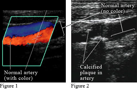
Figure 1 shows a color Doppler ultrasound picture of blood flowing through a normal artery (red) and vein (blue). Figure 2 shows an ultrasound picture of an artery narrowed by the buildup of calcium and fat (cholesterol) in the inner lining of the artery, called plaque, which leads to "hardening of the arteries" (atherosclerosis).
Current as of: October 9, 2017
Author: Healthwise Staff
Medical Review: Kathleen Romito, MD - Family Medicine & Howard Schaff, MD - Diagnostic Radiology