Vision Tests
Test Overview
Vision tests check many different functions of the eye. Some of the tests measure your ability to see details at near and far distances, check for gaps or defects in your field of vision, and evaluate your ability to see different colors. Others may check how sensitive you are to glare (brightness acuity), how well your eyes work together to provide depth perception, and more. Vision tests are usually done along with exams and tests that check the health of the eye. Here are some common tests that check for blurred or low vision.
- Visual acuity (sharpness) tests. These tests help your doctor find out if you have a problem that affects how well you can see. They measure the eye's ability to see details at near and far distances. The tests usually involve reading letters or looking at symbols of different sizes on an eye chart. Usually, each eye is tested by itself. And then both eyes may be tested together, with and without corrective lenses (if you wear them). Several types of visual acuity tests may be used.
- Refraction test. This test shows your level of refractive error and finds out the right prescription for glasses or contact lenses. Refractive errors, such as nearsightedness or farsightedness, occur when light rays entering the eye can't focus exactly on the nerve layer (retina) at the back of the eye. This causes blurred vision. Refraction is done as a routine part of an eye exam for people who already wear glasses or contact lenses. But it will also be done if the results of the other visual acuity tests show that your eyesight is below normal and can be corrected by glasses.
- Visual field tests. They are used to check for gaps in your side (peripheral) vision. Your complete visual field is the entire area seen when your gaze is fixed in one direction. The complete visual field is seen by both eyes at the same time. It includes the central visual field-which detects the highest degree of detail-and the peripheral visual fields.
- Color vision tests. These tests check your ability to distinguish colors. They are used to screen for color blindness in people with suspected retinal or optic nerve disease or who have a family history of color blindness. Color vision tests are also used to screen applicants for jobs in fields where color perception is essential, such as law enforcement, the military, or electronics. Color vision tests only detect a problem-further testing is needed to identify what is causing the problem.
Why It Is Done
Visual acuity tests
These tests may be done:
- As part of a routine eye exam to screen for vision problems. How often you should have routine eye exams changes as you age. Adults and children and teens have different schedules for eye exams.
- To monitor an eye problem, such as diabetic retinopathy, or to find out if a treatment is working.
- To determine if you need glasses or contact lenses to improve your vision.
- After an injury to the eye, to check if your sight was affected.
- When you obtain or renew your driver's license or for some types of employment.
- To check the near vision of school-age children who have trouble reading, poor school performance, or blurred vision while doing work up close.
Refraction
This test is done:
- To determine the correct prescription for eyeglasses or contact lenses.
- To find out if blurred vision is caused by refractive error or eye disease.
Visual field tests
These may be done:
- To check for vision loss in any area of your visual field.
- To screen for eye diseases, such as macular degeneration and glaucoma, which cause gaps in the visual field.
- To look for damage to the nerves of the eye following a stroke, head injury, or other condition that causes reduced blood flow to the brain.
Color vision tests
These tests may be done:
- As part of a routine eye exam.
- To screen for or diagnose color blindness.
- To screen applicants for jobs in which color perception is important, such as truck driving, electronics, or the military.
How To Prepare
If you wear glasses or contact lenses, bring them with you to the exam since the tests cannot be properly performed without them. If you have a copy of your current eyeglass prescription, bring it with you.
If you have a young child, it is best to practice eye tests at home before you take your child to the appointment. This can help your child cooperate better during the real testing. For more information, see the topic Pediatric Preparation for Medical Tests.
Many medicines may affect the results of vision tests. Be sure to tell your doctor about all the over-the-counter and prescription medicines you take.
Talk to your doctor about any concerns you have regarding the need for vision tests, how they will be done, or what the results will mean. To help you understand the importance of these tests, fill out the medical test information form (What is a PDF document?).
How It Is Done
Visual acuity testing
Visual acuity tests are used to evaluate eyesight. Several types of visual acuity tests may be used.
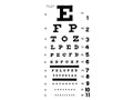
- The Snellen testchecks your ability to see at distances. It uses a wall chart that has several rows of letters. The letters on the top row are the largest; those on the bottom row are the smallest.
- You will stand or sit 20 ft (6 m) from the chart and be asked to cover one eye and then read the smallest row of letters you can see on the chart. If you are unable to cover your eye, an eye patch will be placed over your eye.
- Each eye is tested separately. You may be given a different chart or asked to read a row backward to make sure that you did not memorize the sequence of letters from the previous test.
- If you wear glasses or contacts, you may be asked to repeat the test on each eye while wearing them.
- Let your doctor know if you have trouble reading the letters on one side of the row, or if some letters disappear while you are looking at other letters. You may have a visual field problem, and visual field tests may be needed.
- The E chart tests the vision of children and people who cannot read. The E chart is similar to the Snellen chart in that there are several rows, but all of the rows contain only the letter E in different positions. The top row is the largest and the bottom row of Es is the smallest. You will be asked to point in the same direction as the lines of the E. Similar charts use the letter C or pictures. These charts are also available in a handheld card.
- The Near test uses a small card (Jaeger chart) containing a few short lines or paragraphs of printed text to test your near vision. The size of the print gradually gets smaller. You will be asked to hold the card about14 in. (36 cm) from your face and read aloud the paragraph containing the smallest print you can comfortably read. Both eyes are tested together, with and without corrective lenses. This test is routinely done after age 40, because near vision tends to decline as you age (presbyopia).
If you cannot read any of the letters or print on these charts because of poor vision, your visual acuity will be tested by other techniques, such as counting fingers, detecting hand movements, or distinguishing the direction or perception of light sources (such as room light or a penlight held up close to the face).
Visual acuity tests usually take about 5 to 10 minutes. They may be performed by a nurse, a medical assistant, an ophthalmologist, an optometrist, a teacher, or some other trained person. Testing may be done at a doctor's office, school, workplace, health fair, or elsewhere.
Refraction
Refraction is a test that measures the eye's need for a corrective lens (refractive error). For this test, you will be asked to describe the effects of looking at an eye chart through various corrective lenses.
Your doctor may use eyedrops to widen (dilate) your pupils before you start this test. The eyedrops take about 15 to 20 minutes to dilate the pupil fully.
The doctor may put a device (called a phoropter) in front of your eyes. The device contains many different lenses. Testing one eye at a time, the doctor will ask you to compare the effects of two lenses (first one lens, then the other). You should state which lens of each pair gives you better vision. The doctor will continue to test your eyes with different lenses until it is determined which lenses correct your vision the best. Your doctor may use a hand-held device (retinoscope) to shine light into your eyes. A series of trial lenses will be placed in front of your eyes and adjusted until the light rays are properly focused on your retina.
Visual field tests
Visual field tests are used to check for gaps in your range of vision. They can help detect eye diseases or nervous system problems that limit your ability to see objects clearly in the entire visual field or in one part of it. Several tests are commonly done to evaluate a person's visual field.
- The confrontation test. Your doctor sits or stands2 ft (0.6 m) to3 ft (1 m) in front of you. You cover one eye while fixing your gaze on his or her nose. He or she slowly moves a finger or hand from the outer edge of your visual field toward the center and from the center toward the edge through all areas of your visual field. You will focus your eye on your doctor's nose and signal when you first see his or her finger or hand. The test is then repeated for the other eye.
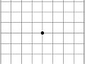
- The Amsler grid test checks for macular degeneration, a disease that causes loss of vision in the center of your visual field. The test uses a4 in. (10 cm) square chart with straight lines that form boxes. The grid has a black dot at the center. The chart is held about14 in. (36 cm) from your face. You cover one eye while focusing your other eye on the black dot. The test is then repeated on the other eye. Tell your doctor if:
- You cannot see the black dot.
- You see a blank or dark spot (other than the center dot).
- The lines in the grid look wavy, blurred, or curved instead of straight. You will be asked to point to the specific abnormal area of the grid.
- Perimetry testing uses a machine that flashes lights randomly at various points in the visual field. You look inside a bowl-shaped instrument called a perimeter. While you stare at the center, lights will flash, and you press a button each time you see a flash. A computer records the location of each flash and whether you pressed the button when the light flashed in that location. At the end of the test, a printout shows any areas of your visual field where you did not see the flashes of light. In an alternative manual perimetry test, your doctor moves a light target and notes your visual field on paper.
- Thetangent screen test uses a black screen with concentric circles and lines leading out from a center point (like a bull's-eye). Sitting3 ft (1 m) to6 ft (2 m) away from the screen, you cover one eye while fixing your gaze on a target point marked on the screen. Test objects of various sizes at the tip of a wand are then moved inward from the outer edge of the screen toward the center. You will signal when you can see the object, and that point is then marked on the screen. The points on the screen where you see the objects are connected to provide an outline of your visual field. The test is then repeated for the other eye. An alternative manual tangent screen test uses a white object against a black background. If you wear glasses, you will keep them on for this test.
Color vision tests
Color vision tests check your ability to distinguish colors. In the most commonly used color vision test, you look for different colored numbers or symbols hidden in varying backgrounds of colored dots.
First, you are shown sample patterns and told what symbols and numbers you can expect to see. You then sit at a table and cover one eye. The doctor holds the color test patterns about 14 in. (36 cm) away from you. Some patterns are harder to pick out than others. As the doctor holds up a pattern, you will identify the number or symbol you see and trace it using a pointer. Some patterns may not have a number or symbol. The test is then repeated with the other eye.
How It Feels
You should not feel any discomfort during these vision tests.
When dilating eyedrops are used
Dilating drops may make your eyes sting and cause a medicine taste in your mouth. You will have trouble focusing your eyes for up to 12 hours after your eyes have been dilated. Your distance vision usually is not affected as much as your near vision, though your eyes may be very sensitive to light. Do not drive for several hours after your eyes have been dilated, unless your doctor says it is okay. Wearing sunglasses may make you more comfortable until the effect of the drops wears off.
Risks
In some people, the dilating eyedrops can cause an allergic reaction.
Results
Vision tests check many different functions of the eye. Your doctor will let you know if your eyesight is normal or if it is better or worse than normal. He or she may also be able to tell you why you have a vision problem.
Visual acuity testing
The visual acuity score compares your distance vision with that of people who have normal vision, using an eye chart. Each eye's score is expressed as two numbers, such as 20/20 (6/6) or 20/100 (6/30). The first number is the distance you stand from the chart, usually 20 ft (6 m) when using a typical wall chart. The second number is the distance from which people with normal eyesight can read the same line on the eye chart.
20/20 (6/6) vision is considered normal. A person with 20/20 vision can see at 20 ft (6 m) what people with normal vision can see at this distance.
- When the second number is smaller than the first number, the person's vision is better than normal. For instance, a person with 20/10 (6/3) vision can see from20 ft (6 m) what people with normal vision can see from10 ft (3 m).
- When the second number is larger than the first number, the person's distance vision is worse than normal.
- A person with 20/200 (6/60) vision or less in his or her best eye when wearing corrective lenses is considered legally blind.
Your doctor will also tell you if you have reduced near vision.
Refraction
The doctor tests your eyes with different lenses until the lens that corrects your vision the best (sometimes better than 20/20 or 6/6) is found. The result of a refraction test determines your prescription eyeglass or contact lens strength.
Visual field test
Normally, a person's visual field forms a rough circle with a natural blind spot. If your vision is normal, you should be able to see objects clearly throughout the entire visual field except for the area with the natural blind spot. When you are using both eyes to see, the blind spots do not interfere with your vision.
You may have vision loss in certain areas of the visual field if you are not able to see:
- Test objects during tangent screen testing.
- Movements or light flashes during perimetry testing.
Abnormal results during Amsler grid testing include:
- Not being able to see the black dot at the center of the grid.
- Not being able to see all four edges of the grid.
- Having blank spots or dark spots on the grid (other than the black dot at the center).
- Seeing lines that look wavy or curved.
Gaps in different parts of the visual field may have many causes, including eye diseases (such as glaucoma and macular degeneration) or nervous system problems (such as stroke). If results on any of the visual field tests are abnormal, you will need further tests to determine the cause.
Color vision test
People who have normal color vision are able to distinguish the colored numbers, symbols, or paths from the background of colored dots.
If you are not able to distinguish some or all of the colored patterns from the background, you may have a color vision problem. You may be able to pick out some patterns of colors but not others. Or you may be able to pick out patterns that are different from a person with normal vision, depending on what type of color vision problem you have.
Many conditions can change your vision test results. Your doctor will discuss any significant abnormal results with you in relation to your symptoms and past health.
What Affects the Test
Reasons you may not be able to have the test or why the results may not be helpful include:
- Your ability to understand or follow instructions. Some vision tests cannot be done on babies, small children, or people who cannot understand or follow the instructions.
- Your ability to stay alert and respond to questions.
- Failure to wear prescribed eyeglasses or contact lenses.
- Poor lighting.
What To Think About
- A complete eye and vision evaluation also includes a physical exam of the structures inside the eye. To learn more, see the topic Ophthalmoscopy.
- A test to screen for increased intraocular pressure (IOP), which increases your risk for glaucoma, is often part of a routine eye exam. It also is used to monitor treatment for glaucoma. Tonometry can be used to determine whether a medicine is keeping your IOP below a set target pressure determined by your doctor. To learn more, see the topic Tonometry.
- Home tests for near vision in adults and distance vision in children are available. These tests should not replace a thorough eye exam by a doctor.
- Until your child's visual system has fully developed, the doctor may check your child's visual behavior. This includes testing eye movements, the alignment of the eyes, how well your child can track an object, whether your child has depth perception, and how well the two eyes work together. If you notice signs of eye problems in your child, talk to your child's doctor. To help you decide if you need to see a doctor for a vision problem, see the topic Eye Problems, Noninjury.
References
Other Works Consulted
- American Academy of Ophthalmology (2012). Refractive Errors and Refractive Surgery (Preferred Practice Pattern). San Francisco: American Academy of Ophthalmology. Also available online: http://one.aao.org/CE/PracticeGuidelines/PPP_Content.aspx?cid=0bc8c7ce-26df-46da-bf2b-7e908bedaf64.
- Chang DF (2011). Ophthalmologic examinations. In P Riordan-Eva, ET Cunningham, eds., Vaughan and Asbury's General Ophthalmology, 18th ed., pp. 27-57. New York: McGraw-Hill.
- Chernecky CC, Berger BJ (2013). Laboratory Tests and Diagnostic Procedures, 6th ed. St. Louis: Saunders.
- Fischbach FT, Dunning MB III, eds. (2009). Manual of Laboratory and Diagnostic Tests, 8th ed. Philadelphia: Lippincott Williams and Wilkins.
Credits
ByHealthwise Staff
Primary Medical Reviewer Kathleen Romito, MD - Family Medicine
Adam Husney, MD - Family Medicine
E. Gregory Thompson, MD - Internal Medicine
Current as ofDecember 3, 2017
- Top of Page
Next Section:
Why It Is Done
Previous Section:
Test Overview- Top of Page
Next Section:
How To Prepare
Previous Section:
Why It Is Done- Top of Page
Next Section:
How It Is Done
Previous Section:
How To Prepare- Top of Page
Next Section:
How It Feels
Previous Section:
How It Is Done- Top of Page
Next Section:
Risks
Previous Section:
How It Feels- Top of Page
Next Section:
Results
Previous Section:
Risks- Top of Page
Next Section:
What Affects the Test
Previous Section:
Results- Top of Page
Next Section:
What To Think About
Previous Section:
What Affects the Test- Top of Page
Next Section:
References
Previous Section:
What To Think About- Top of Page
Next Section:
Credits
Previous Section:
References- Top of Page
Current as of: December 3, 2017